How is Waldenstrom macroglobulinemia diagnosed?
Waldenstrom macroglobulinemia (WM) is often discovered incidentally when a person gets blood tests done for some other reason or during their annual check-up. Sometimes, people see their doctor complaining of specific symptoms or have a general sense of not feeling well. If signs and symptoms suggest that a person might have WM, a number of tests and exams will be performed to confirm, or rule out, a diagnosis of WM.
Health history and physical exam
Your doctor will start by getting a detailed medical history. He or she will ask questions about your past and current health, including:
- Any symptoms you are experiencing, particularly those that may be related to WM
- Any illnesses or surgeries you have had
- What medications you take, including prescription and over-the-counter medicines
- What your current lifestyle is like (exercise, diet, tobacco or alcohol use, hobbies, etc.)
- Your family medical history
Next, your doctor will perform a physical exam, paying special attention to areas of your body that might be involved with WM, like your lymph nodes, spleen, liver and eyes. Your doctor will also order a number of tests.
Blood tests
- Your doctor will order several blood tests to determine the types and amounts of antibodies in your blood. People with WM have a high level of the antibody IgM (immunoglobulin M). These blood tests are called:
- Serum protein electrophoresis
- Serum immunofixation electrophoresis
- Serum quantitative immunoglobulins
- A complete blood count (CBC) is a commonly used blood test. It measures the levels of red blood cells, white blood cells (including lymphocytes), and platelets. Because WM cells may crowd out the production of healthy blood cells in the bone marrow, people with WM often have low blood counts.
- A comprehensive metabolic panel (CMP) is a panel of blood tests used to determine how well your organs – such as your kidneys and liver – are working.
- There are other blood tests used to monitor WM, detect complications of the disease, and help predict a person’s prognosis:
- Serum viscosity: Measures the thickness (viscosity) of the blood. In WM, high levels of IgM in the blood make it thicker than normal.
- Cryocrit and cold agglutinins: These two tests measure for certain abnormal IgM antibodies seen in some people with WM. These antibodies react to cold temperatures and can affect the level of IgM in the blood.
- Beta-2 microglobulin: Measures the blood level of a protein called beta-2 microglobulin. It provides information about the severity of the disease; high levels are linked with a worse outlook.
Bone marrow aspiration and biopsy
Since the symptoms of WM can be similar to those caused by other diseases or infections, a diagnosis of WM can only be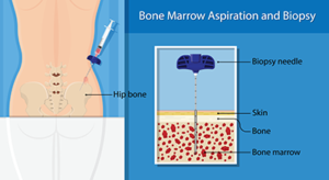 confirmed by testing your bone marrow. The bone marrow samples are sent to a laboratory to be examined by an expert in diagnosing diseases of the bone marrow and blood, called a hematopathologist. He or she will look for the lymphoplasmacytic cells of WM, which have features of both B-lymphocytes and plasma cells.
If detected, advanced analysis of the bone marrow tissue can further characterize the cells’ characteristics, including their genetic mutations. There are two gene mutations that may be found in people with WM: Most have an abnormal (mutated) version of a gene called MYD88; about 40 percent of people with WM have a mutation of the gene CXCR4; 5% percent will not have a mutation of either of them.
Bone marrow aspiration and biopsy can be done at your doctor’s office or in the hospital. These procedures are performed at the same time, usually on the back of your pelvic bone. Your doctor will numb the area with a local anesthetic and insert a needle to remove liquid bone marrow with a syringe (aspiration). Then a wider needle will be inserted to remove a small piece of bone and soft marrow (biopsy). You may experience some brief pain, tenderness, or bruising at the biopsy site. Most people go home following the procedure.
Imaging tests
Imaging tests use x-rays, sound waves, magnetic fields, or radioactive particles to produce detailed pictures of the inside of your body. They are not needed to diagnose WM; however, they help your doctor determine whether the cancer has spread beyond your bone marrow to other parts of your body.
The most widely used imaging test for WM is computed tomography (CT). CT scans are useful for detecting signs of cancer in your chest, abdomen , back and pelvis. The scan will show if your lymph nodes, spleen, or other organs are enlarged. Sometimes this is done in combination with a positron emission tomography (PET) scan. A PET scan uses a weak radioactive material, which is helpful in detecting small collections of cancer cells, even if the area looks normal on a CT scan. A magnetic resonance imaging scan (MRI) is used predominantly for the brain and spinal cord.
For a detailed description of the medical tests typically used to diagnose and monitor WM, you can refer to the IWMF booklet, Medical Tests, found in IWMF and Affiliate Publications. This booklet can be particularly useful in helping you understand the implications of your test results.


